
In modern dentistry, capturing accurate impressions of the oral cavity has evolved into a swift, precise, and comfortable digital process. An intraoral scanner is a game‑changing device that transforms traditional impression-taking into a seamless, patient-friendly digital workflow. As Iran’s trusted provider of dental imaging tools, Hamin Dental offers expert insights and reliable solutions tailored for today’s dental practices. In this comprehensive guide, we’ll explore intraoral scanners in depth—understanding what they are, how they work, how they benefit patients and dentists, and how to choose the right model for your clinic.

Understanding the Intraoral Scanner
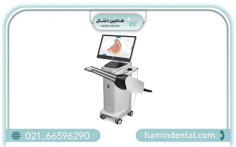
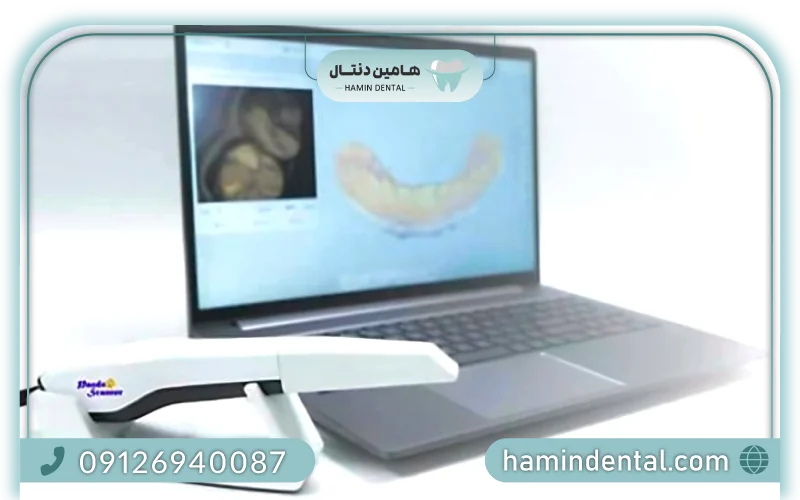
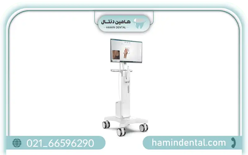
An intraoral scanner is a handheld digital device used by dentists to capture a 3D image of a patient’s teeth, soft tissues, and surrounding oral structures. Instead of traditional impression materials, this scanner projects structured light or laser onto dental surfaces, collects optical data, and constructs a high-resolution digital model in real time. This 3D digital model can be rotated, zoomed, edited, and immediately shared with labs or colleagues
These scanners provide a faster, cleaner, and more accurate method to obtain impressions, replacing trays filled with putty that can feel uncomfortable for patients ([turn0search4]).
Why Intraoral Scanners Matter
Improved Accuracy and Fit
Digital impressions drastically reduce material distortion common in traditional methods, resulting in better-fitting crowns, bridges, dentures, and splints. This higher precision cuts down on remakes and patient reappointments ([turn0search16]).
Patient Comfort and Acceptance
Patients often prefer intraoral scans over conventional impressions, since the scanner is non-invasive, fast, and avoids the unpleasant sensation of bulky trays. This improves overall satisfaction and compliance ([turn0search4]).
Time Efficiency and Workflow
Scans are captured digitally and can be instantly transmitted to a dental laboratory, eliminating time-consuming steps like disinfecting, shipping, and manual model fabrication. This speeds up treatment planning and prosthetic design ([turn0search22]).
Enhanced Communication
3D digital models help dentists clearly explain conditions and treatment options to patients. Visualizing the oral anatomy on a monitor promotes understanding and increases acceptance of recommended treatments.
Sustainability
Digital impressions eliminate the need for impression trays and materials, reducing waste and supporting eco-friendly Dental imaging equipment .
How an Intraoral Scanner Works
-
Scanning Process
A dentist or assistant gently sweeps the small wand over teeth and soft tissues, while onboard LEDs illuminate the area. The device captures multiple images per second, stitching them together into a continuous 3D model.
2. Data Processing
Captured images are converted into point clouds and then triangulated into a mesh model. Advanced software refines and validates scan quality in real time, alerting users to any missing data or errors ([turn0search17]).
3. Editing and Validation
Clinicians can manipulate the scan on screen—correcting imperfections or trimming excess scan information. Some scanners automatically detect incomplete scans to ensure full digital coverage.
4. CAD/CAM Integration
Once finalized, the digital model is sent to CAD software for designing restorations like crowns, bridges, aligners, or surgical guides. It can also be transmitted directly to a manufacturing or milling system ([turn0search21]).
Main Benefits of Digital Scanning
Patient Experience
-
No gag‑inducing trays or messy materials.
-
Fast, non-invasive scanning.
-
Instant visualization builds trust and understanding.
Clinical Accuracy
-
Reduces distortions common in physical impressions.
-
Dramatically decreases fitting issues—remake rates drop by 30–40%.
-
Consistent, repeatable scans across patients and visits.
Operational Efficiency
-
Instant file sharing with labs or interdisciplinary teams.
-
Shorter treatment cycles and fewer appointments.
-
Saves time on disinfecting and shipping traditional impressions.
Cost Savings Over Time
-
No recurring expense for impression materials.
-
Less downtime and fewer case remakes.
-
Streamlined workflows yield better return on investment .
Clinical Applications by Specialty
Prosthodontics
Scans capture fine detail of crown and bridge preparations, enabling precise restorations. Scan bodies can accurately register implant locations. Models may also be used to design partial dentures and smile simulations.
Orthodontics
Used to create digital models for clear aligners, retainers, and bracket setups. Enables monitoring of tooth movement during treatment progress. Special orthodontic workflows integrate IOS data into aligner planning.
Endodontics
Scans support visualization of tooth morphology, crown margins, and root anatomy. When combined with CBCT data, they enhance diagnosis and planning for canal treatments or restorations.
Periodontics
3D models assist in assessing gingival contours, recession, pocket depths, and planning soft tissue procedures. Scans can be tracked over time to monitor periodontal health changes.
Forensic Applications
Scanning of palatal rugae offers a unique biometric record. Studies suggest intraoral scans may support forensic identification due to the stability of palatal morphology over time ([turn0search5]).
Challenges and Limitations
While intraoral scanners offer impressive advantages, some considerations remain:
-
Cost and Learning Curve: Initial investment and training can be significant for smaller clinics ([turn0search17]).
-
Deep Margin Capture: Scanning below the gingival margin can be difficult, especially in bleeding or inflamed tissues.
-
Soft Tissue Distortion: Movements or saliva may impact scan accuracy.
-
Lab Compatibility: Not all labs accept digital workflows yet, which may require coordination ([turn0search14]).
Market Growth and Trends
The global market for intraoral scanners is expanding rapidly. Valued at about USD 347 million in 2024, it is expected to reach USD 764 million by 2032 with strong annual growth (~10% CAGR) ([turn0search15]). Advances like AI-guided scanning, clinic-lab integration, and portable handheld models are accelerating adoption.
Increasingly, scanners integrate with AI to detect anomalies (like caries or cracks), guiding clinicians during acquisition. Cloud-based workflows and remote collaboration tools are also becoming more common
Why Choose Hamin Dental
Hamin Dental serves as an expert source and supplier for intraoral scanners in Iran. Our offerings are selected based on clinical accuracy, user-friendliness, and software compatibility. Choosing us ensures:
-
Complete Product Range, from ergonomic intraoral wands to integrated clinic systems.
-
Expert Guidance to match scanner models with clinical needs and budgets.
-
Training & Support, including onboarding, software training, and technical service.
-
Flexible Options: new systems and certified pre-owned units.
-
Seamless Workflow Integration into clinics using digital software and CAD/CAM systems.
Buying Guide: What to Look For
Scanner Accuracy & Speed
Choose devices with high cutoff resolution and fast capture rates that reduce scanning time and motion artifacts.
Ergonomic Design & Hygiene
Lightweight, ergonomic wands improve operator comfort. Removable, autoclavable sleeves ensure infection control.
Software Features
Look for real-time validation, scan completion alerts, and easy export to CAD/Dental labs.
Compatibility
Ensure integration with digital lab systems and file formats (STL, PLY) for smooth transfer.
Training & Maintenance
Opt for suppliers that provide staff training, software updates, and local technical support.
Warranty & Upgrade Paths
Look for clear warranty terms and future software or hardware upgrade possibilities.
Clinical Workflow and Deployment
Embedding an intraoral scanner into everyday dental workflows often follows this process:
-
Pre-Scan Preparation: Clean teeth and tissues, retract lips/tongue.
-
Scanning the Arch: Sweep the wand over the arch, capturing occlusion and anatomy.
-
Model Validation: Review real-time 3D model, correct missing areas immediately.
-
Export / Send to Lab: Share digital files with CAD technicians or milling centers.
-
Patient Consultation: Use the 3D model to explain treatment options.
-
Case Refinement: Architect crowns, bridges, appliances with the scan data.
-
This streamlined process enhances patient service, reduces errors, and accelerates production timelines.
Return on Investment (ROI) in Practice
Though scanners have upfront costs, ROI becomes clear through:
-
Fewer repeated impressions and remakes.
-
Reduced use of impression materials.
-
Time savings for both clinicians and lab technicians.
-
Faster case turnaround and increased patient throughput.
-
Improved patient satisfaction and treatment acceptance rates.
Over time, clinics equipped with intraoral scanners often report lower material costs, higher productivity, and more positive reviews among patients.
Future of Intraoral Scanning
The future of IOS technology is bright. Look for:
-
AI‑guided scans that flag anomalies and sequencing errors.
-
Wireless handheld units for improved ease and portability.
-
Cloud‑based collaboration tools for seamless communication with labs and specialists.
-
Augmented Reality (AR) integration for real-time treatment planning.
-
Enhanced resolution and subgingival capture for deeper margin detection.
-
Clinics partnering with Hamin Dental can access these forward-looking tools as they become clinically available.
Frequently Asked Questions
Q1: How long does a typical scan take?
Most full-arch scans complete within 1–3 minutes, depending on operator speed and scanner model.
Q2: Can intraoral scanners replace traditional impressions entirely?
For most restorative, orthodontic, and implant work, yes—they are accurate enough for final fabrication. In rare cases of subgingival margins or soft tissue restorations, physical impressions may still be needed.
Q3: How accurate are scan-based restorations compared to analog?
Studies show intraoral scanners deliver superior margin fit and reduced internal discrepancies for crowns and bridges ([turn0search7]).
Q4: Are intraoral scanners hygienic?
Yes—wands use disposable or autoclavable barriers and are easy to disinfect between patients.
Q5: Is there a learning curve for staff?
Some initial training is required, but most find the scanner intuitive. Support and training from suppliers accelerate adoption.